MIDDLE EAR INFECTIONS
Middle ear infections, also known as otitis media, are a common ailment that can affect individuals of all ages, although they are particularly prevalent among children.
The symptoms of a middle ear infection can range from mild to severe and may include ear pain, fever, and hearing difficulties. Recognising the debilitating impact such conditions can have on a patient’s quality of life, Dr Sandeep prioritises prompt and accurate diagnosis.
ACUTE OTITIS MEDIA (AOM)
Acute Otitis Media (AOM) is a type of middle ear infection that is characterised by the sudden onset of symptoms such as ear pain, fever, and sometimes hearing loss. It is a condition primarily affecting the middle ear, which is the air-filled space located behind the eardrum that contains the tiny vibrating bones of the ear. AOM is most commonly caused by bacterial or viral infections and is particularly prevalent among children, although it can occur at any age.
Clinical Features
In AOM, the middle ear becomes inflamed and filled with pus or fluid due to an infection. The following are common symptoms:
- Severe ear pain
- Fever
- Irritability or fussiness (commonly observed in children)
- Hearing difficulties
- Fluid drainage from the ear
How It Differs from Otitis Media with Effusion
It’s important to distinguish AOM from Otitis Media with Effusion (OME), another type of middle ear condition. While AOM is an active infection with symptoms like pain and fever, OME is a condition where fluid remains trapped in the middle ear but without the acute symptoms of infection. OME often follows an episode of AOM but can also occur independently.
DIAGNOSIS
At The ENT Clinic, Dr Sandeep Uppal takes a meticulous approach to the diagnosis of Acute Otitis Media (AOM). Understanding that accurate diagnosis is the cornerstone of effective treatment, our team employs a multi-faceted methodology:
Clinical Interview
The first step often involves a detailed clinical interview where the patient or the caregivers are asked about the onset, duration, and nature of symptoms. Questions may include:
- When did the ear pain start?
- Are there any accompanying symptoms like fever or hearing loss?
- Is there a history of upper respiratory infections or allergies?
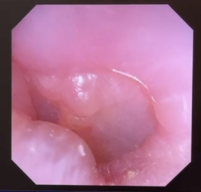
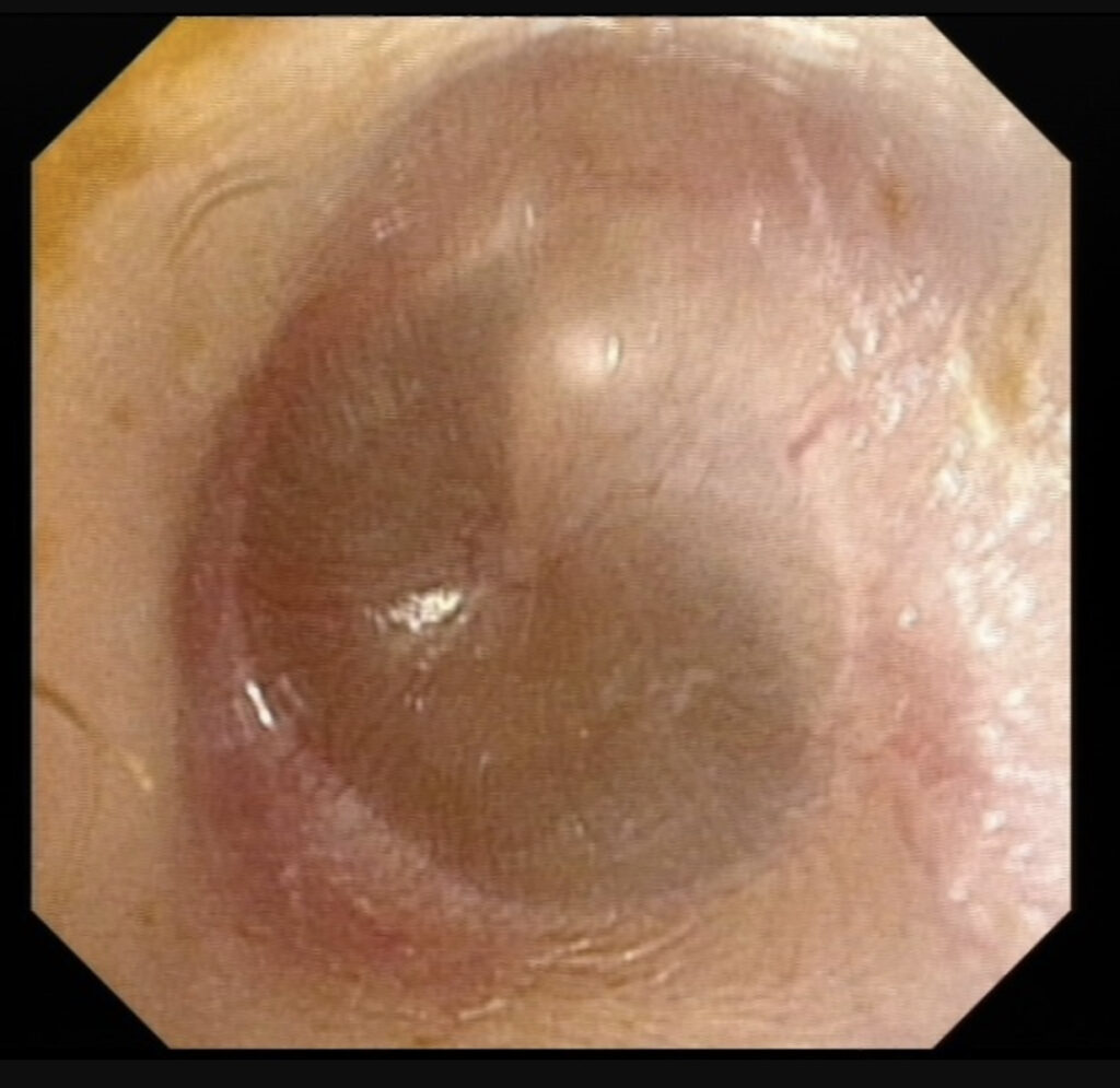
Otoscopic Examination
An otoscope, a medical device equipped with a light and a magnifying lens, is used to closely examine the ear canal and eardrum. The team looks for:
- Redness and inflammation of the eardrum
- Presence of fluid or pus behind the eardrum
- Eardrum immobility
- Any perforation or rupture of the eardrum
Tympanometry
This diagnostic test measures the movement of the eardrum in response to air pressure changes. Lack of normal eardrum movement usually indicates the presence of fluid in the middle ear, which is a hallmark of AOM.
Pneumatic Otoscopy
This advanced form of otoscopy allows the practitioner to not only visualise the eardrum but also to assess its mobility by blowing a small puff of air into the ear canal. Reduced or absent movement of the eardrum suggests fluid accumulation and middle ear infection.
Audiometry
In cases where hearing loss is reported or suspected, an audiometric test might be conducted to assess the extent of hearing impairment.
Microbiological Tests
Although less common in routine practice for AOM, swabs for microbiological culture may be taken in recurrent or persistent cases to identify the specific bacterial or viral pathogen responsible for the infection.
Through a combination of these diagnostic tools and methods, we aim to provide a comprehensive and accurate diagnosis of Acute Otitis Media. This thorough diagnostic process is essential for devising an effective treatment plan tailored to the needs of each individual patient.
TREATMENT
At The ENT Clinic, Dr Sandeep and his team are adept at offering evidence-based treatments for Acute Otitis Media (AOM), customising the approach based on the severity of the condition, the age of the patient, and any accompanying medical concerns. Here’s a closer look at their treatment modalities:
Antibiotic Therapy
For bacterial AOM, antibiotics are often the first line of treatment. The choice of antibiotic may vary based on the suspected causative agent and the patient’s medical history.
Pain Management
Symptomatic relief is crucial in managing AOM, especially in the initial stages when pain can be intense. Analgesics such as paracetamol or ibuprofen are commonly prescribed for pain and fever.
Observation
In certain cases, especially when the infection is suspected to be viral or when symptoms are mild, a “watchful waiting” approach may be adopted. This involves monitoring symptoms closely for 48 to 72 hours and initiating antibiotic treatment only if there is no improvement.
Myringotomy
In severe or recurrent cases of AOM where fluid accumulation is significant, a surgical procedure known as myringotomy may be considered. This involves making a small incision in the eardrum to allow drainage of the trapped fluid.
Tympanostomy Tubes
For chronic or recurrent AOM, the insertion of tympanostomy tubes (also known as grommets) may be recommended. These tubes help to equalise pressure and allow fluid to drain from the middle ear, thereby reducing the frequency of infections.
Adjunct Treatments
In some cases, adjunct treatments like decongestants or antihistamines may be recommended, especially if the patient has concurrent issues like allergies or sinusitis that could be exacerbating the condition.
Follow-up and Monitoring
We place a strong emphasis on follow-up care to monitor the patient’s response to treatment. This often involves another otoscopic examination and possibly repeat tympanometry to confirm that the infection and any fluid accumulation have resolved.
Potential Complications of Acute Otitis Media
Through a comprehensive and individualised treatment plan, Dr Sandeep Uppal and his team at The ENT Clinic aim to manage Acute Otitis Media effectively, alleviating symptoms and preventing complications. Our experienced team are well aware of the potential complications that can arise from untreated or inadequately managed Acute Otitis Media (AOM). Their focus on thorough diagnosis and effective treatment is geared towards minimising these risks. Here are some complications associated with AOM:
Hearing Loss
One of the most immediate complications of untreated AOM is temporary hearing loss. The accumulation of fluid in the middle ear can impede the movement of the eardrum and the auditory ossicles, leading to decreased hearing. While this is often reversible, chronic untreated AOM may result in permanent hearing loss.
Tympanic Membrane Perforation
Persistent infection and fluid accumulation can lead to increased pressure against the eardrum, causing it to rupture or perforate. This usually results in sudden relief from pain but may require surgical repair.
Chronic Otitis Media
If AOM episodes are recurrent or persistent, they can lead to chronic otitis media. This condition is characterised by long-term infection or inflammation of the middle ear and may require more aggressive treatment, including surgery.
Mastoiditis
The mastoid bone, located behind the ear, can become infected if AOM is not adequately treated. Mastoiditis is a severe condition that requires immediate medical intervention, often involving intravenous antibiotics and possibly surgical drainage.
Facial Nerve Paralysis
Though extremely rare, untreated AOM can lead to facial nerve paralysis due to the close anatomical relationship between the facial nerve and the middle ear. If the facial nerve is affected the patient notices partial or total loss of ability to move the face on the side of the affected ear.
Intracranial Complications
In the most severe cases, untreated AOM can lead to intracranial complications like meningitis or a brain abscess. These are medical emergencies requiring immediate hospitalisation and intensive care.
Otitis Media with Effusion
Following an episode of AOM, fluid may remain in the middle ear for several weeks, leading to a condition known as Otitis Media with Effusion (OME). The fluid is often thick and sticky, resembling glue, hence the colloquial term. It’s particularly prevalent among children but can also affect adults. While not an acute infection, OME can still impair hearing and requires monitoring.
Given these potential complications, Dr Sandeep Uppal and his team adopt a proactive approach to the diagnosis and treatment of AOM. Their comprehensive care model ensures that each patient receives individualised treatment aimed at not only resolving the immediate symptoms but also preventing these more severe outcomes. Through timely intervention and expert care, the team at The ENT Clinic aims to mitigate the risks associated with Acute Otitis Media.
GLUE EAR (OTITIS MEDIA WITH EFFUSION)
Otitis Media with Effusion (OME), which is commonly known as “glue ear.” This condition is characterised by the accumulation of fluid in the middle ear behind the eardrum. It’s particularly prevalent among children but can also affect adults. The fluid is often thick and sticky, resembling glue, hence the colloquial term.
Diagnosis
Diagnosing OME usually involves a combination of clinical history, physical examination, and sometimes additional tests:
Clinical History
The patient often presents with hearing difficulties, a feeling of fullness in the ear, and sometimes, balance issues. However, pain is usually not a prominent symptom in OME.
Physical Examination
Dr Sandeep Uppal would typically use an otoscope or a microscope to examine the ear. Signs of fluid or an immobile or retracted eardrum are indicative of OME.
Tympanometry
This test can be performed to assess how well the eardrum is moving and to confirm the presence of fluid in the middle ear.
Audiometry
A hearing test might be recommended to assess the level of hearing loss, if any.
Treatment
Treatment for OME often varies depending on the severity of the condition, patient age, and associated symptoms. Here are some common treatment options:
Watchful Waiting
In many cases, especially in children, the condition resolves on its own. Dr Sandeep and his team may adopt a ‘watch and wait’ approach, monitoring the situation over time.
Medical Treatment
In some cases, a course of nasal decongestants or antihistamines may be prescribed to alleviate symptoms. However, the efficacy of these medications in treating OME is debated.
Ventilation Tubes (Grommets)
For persistent or severe cases, Dr Sandeep may recommend the insertion of ventilation tubes, known as grommets, into the eardrum. This allows the fluid to drain, alleviating symptoms and preventing complications.
Adenoidectomy
Sometimes large adenoids at the back of the nose block the Eustachian tubes, which in turn contributes to glue ear. In such patient’s removal of the adenoids is advised, especially if the patient has recurring episodes of OME and/or other nasal symptoms.
Hearing Aids
In cases where OME leads to significant hearing loss, hearing aids may be recommended to improve auditory function. Often this is a temporary measure to give the ear enough time to recover spontaneously.
POTENTIAL COMPLICATIONS OF OTITIS MEDIA WITH EFFUSION
Dr Sandeep is particularly vigilant when it comes to managing complications that may arise from Otitis Media with Effusion (OME). While OME is often a self-limiting condition that may resolve on its own, there are instances where it can lead to complications. Understanding these potential complications is crucial for providing effective patient care.
Hearing Loss
One of the most immediate complications is a temporary reduction in hearing. This can be particularly problematic for children in developmental stages, affecting speech and academic performance.
Speech and Developmental Delays
In children, prolonged OME and resultant hearing loss can lead to delays in speech development, as well as other developmental milestones.
Balance Issues
The accumulation of fluid in the middle ear can affect the equilibrium, leading to balance problems or dizziness.
Tympanic Membrane Retraction or Atrophy
Over time, the pressure from the accumulated fluid can cause the eardrum to retract or become thin, which could potentially result in permanent damage.
Chronic Suppurative Otitis Media
In some cases, OME can progress to a more severe form of ear infection that involves persistent drainage from the ear.
Cholesteatoma
Rarely, chronic OME can lead to the formation of a cholesteatoma, a skin cyst that can grow and destroy the bones of the middle ear, leading to hearing loss and other serious complications.
Social and Behavioural Impact
Especially in children, recurrent episodes of OME and associated hearing loss can result in behavioural issues or decreased social interaction.
Management of Complications
Given these potential complications, Dr Sandeep Uppal and his team adopt a thorough approach in managing OME. They emphasise early diagnosis and appropriate intervention, which may include surgical options such as the insertion of ventilation tubes (grommets) or adenoidectomy to prevent long-term issues.
Additionally, they may work in collaboration with audiologists for hearing assessments and speech therapists for addressing any developmental delays, providing a multidisciplinary approach to the comprehensive care of OME and its complications.