


Parotid Surgery
PAROTID SURGERY TAKE A CONFIDENT STEP TOWARDS PAROTIDECTOMY: SKILLED INTERVENTION…
Parotid Gand and Duct Stones
PAROTID GAND AND DUCT STONES WHAT YOU MIGHT FEEL PAIN…
Outer Ear Infections
OUTER EAR INFECTIONS Outer ear infections, or swimmer’s ear, can…
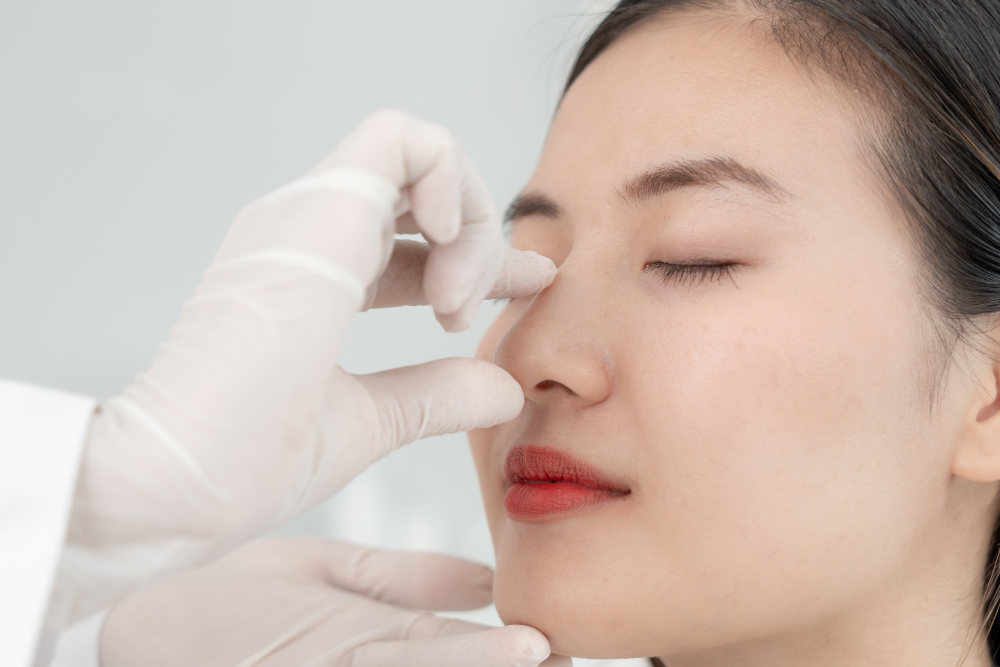
Nasal Foreign Body Management
NASAL FOREIGN BODY MANAGEMENT TYPES OF NASAL FOREIGN BODIES ORGANIC…
Management of Thyroid Lumps
MANAGEMENT OF THYROID LUMPS Dr Sandeep Uppal and his team…
Management of Snoring and Obstructive Sleep Apnoea
MANAGEMENT OF SNORING AND OBSTRUCTIVE SLEEP APNOEA CONSULTATION AND HISTORY…
Management of Sinusitis
MANAGEMENT OF SINUSITIS WHAT CAUSES SINUSITIS? Understanding the root causes…
Management of Enlarged and Infected Adenoids
MANAGEMENT OF ENLARGED AND INFECTED ADENOIDS WHAT ARE ADENOIDS? The…
MALFUNCTION OF THYROID GLAND AND ITS MANAGEMENT
MALFUNCTION OF THYROID GLAND AND ITS MANAGEMENT WHAT HAPPENS WHEN…