OTOPLASTY (PINNAPLASTY)
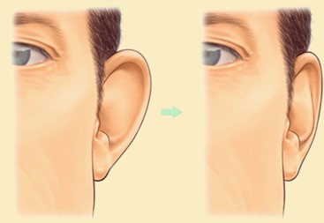
Otoplasty, also known as pinnaplasty, is a surgical procedure designed to reshape or reposition the ears. It’s commonly performed to correct prominent or protruding ears but can also address other ear deformities. The goal is to produce a more natural appearance and to improve the balance and proportion of the ears in relation to the face.
Here are some key points about Otoplasty:
INDICATIONS
The most common reason people seek Pinnaplasty is for prominent ears, but it can also be used to correct other ear deformities or to repair damage from injuries.
WHAT IS THE BEST TIME TO HAVE OTOPLASTY?
The best time for surgery can depend on several factors:
RECOMMENDED AGE FOR SURGERY
While the procedure can be performed on adults, it’s often done on children between the ages of 5 and 14. By age 5, the ear is typically fully grown, so the results of the surgery will be long-lasting.
Performing the surgery at a younger age before starting formal schooling can also prevent potential teasing or self-consciousness. It is recommended to perform the procedure before the child starts school although it can be done in later years.
With age the ear cartilage often undergoes calcification and becomes rigid. In certain older individuals, the extent of this calcification can make otoplasty more difficult and less effective, with a higher risk of the ear reverting to its original protruding state.
EMOTIONAL READINESS
It is important for the client, whether a child or an adult, to be emotionally prepared for the surgery and to have realistic expectations about the results.
PHYSICAL HEALTH
Good overall health is important for any surgical procedure, including Otoplasty. Clients should be free from any major health issues that could affect surgery or recovery.
TIMING IN LIFE
For adults, scheduling the surgery may depend on personal and professional commitments. They need to consider the recovery period and whether they can take time off from work or social activities.
SEASONAL CONSIDERATIONS
Some clients may prefer to schedule the surgery during cooler months when it’s easier to wear headbands or dressings without discomfort, or when they have a break from school or work, such as during the winter or summer holidays.
Ultimately, the best time for Pinnaplasty is a personal decision that should be made in consultation with a qualified surgeon like Dr Sandeep, considering the client’s unique circumstances, including their physical and emotional readiness, as well as practical considerations for recovery.
OTOPLASTY PROCEDURE
The specific techniques used can vary based on the individual’s needs. The procedure may be performed by making an incision either behind or in front of the ear.
The cartilage is then reshaped, and sometimes non-removable stitches are used to help maintain the new shape. Excess skin or cartilage may be removed if necessary.
Here is a general outline of how the Otoplasty procedure is typically performed:
PREOPERATIVE EVALUATION
CONSULTATION
An initial consultation with Dr Sandeep will give you ample time to discuss your goals, expectations, potential outcomes, recovery and potential risks in detail.
MEDICAL EVALUATION
Dr Sandeep and his team will carry out a thorough assessment of your medical history. He will then examine the ears to characterize the deformity and suggest a personalized surgical plan. Pre-operative photographs will be taken for documentation and discussion.
ANAESTHESIA
Dr Sandeep and his team ensure that their client is comfortable throughout the procedure. Local anaesthesia with sedation is often used for adults, allowing the patient to be relaxed and pain-free. General anaesthesia administered by an experienced Senior Consultant anaesthetist is the default option in children to ensure they remain asleep throughout the procedure. Many adults prefer to have a general anaesthesia as well.
POSTERIOR TECHNIQUES FOR OTOPLASTY
MUSTARDÉ TECHNIQUE
OBJECTIVE
The primary goal of the Mustardé technique is to recreate or enhance the antihelical fold, which is often underdeveloped or absent in people with prominent ears. The antihelical fold is the inner ridge of the ear that runs parallel to the outer rim (helix). By enhancing this fold, the ear can be positioned closer to the head, reducing its prominence.
PROCEDURE
- An incision is made behind the ear to expose the cartilage.
- Permanent, non-absorbable sutures are placed through the cartilage to create or enhance the antihelical fold.
- The sutures are strategically positioned to reshape the cartilage without removing any part of it.
- If needed a part of the conchal bowl cartilage may be excised or repositioned if it is also contributing to the prominent ear deformity.
- Once the desired shape is achieved, the incision is closed.
ADVANTAGES
- The Mustardé technique is less invasive than some other methods because it doesn’t involve removing cartilage.
- The use of permanent sutures provides lasting results.
POTENTIAL RISKS AND COMPLICATIONS
The Mustardé technique, while effective for correcting prominent ears, does come with potential risks, as does any surgical procedure. Here are some of the potential risks and complications associated with the Mustardé technique:
- Asymmetry: One ear may appear differently than the other in terms of shape, size, or position.
- Recurrence: The ear might gradually return to its previous position or shape over time, especially if the cartilage is resistant to reshaping or if the sutures fail.
- Unsatisfactory Cosmetic Result: Despite the surgery, the client might not be satisfied with the appearance of the ears.
- Over-correction: The ear might be pinned back too closely to the head, resulting in an unnatural appearance.
- Under-correction: The desired correction might not be achieved, and the ear may still appear more prominent than desired. Correction may require further surgery.
- Suture Visibility or Extrusion: The permanent sutures used to reshape the cartilage might be visible through the thin skin of the pinna and may work their way to the surface and become exposed. This can require removal or replacement of the suture.
- Infection: As with any surgical procedure, there’s a risk of infection at the incision site or deeper within the ear. Infection of the ear cartilage has the potential to distort the ear (cauliflower ear) permanently. Antibiotics used at the time of surgery and in the post-operative period help to greatly reduce the risk of infection.
- Scarring: Scarring can occur at the incision site. While the incision is typically made behind the ear and is less visible, some individuals might experience hypertrophic or keloid scarring.
- Hematoma: Blood can accumulate at the surgical site, leading to pain and swelling. This might require drainage.
- Altered Sensation: There might be temporary or permanent changes in skin sensation around the operated area, including numbness or increased sensitivity.
- Adverse Reaction to Anaesthesia: If general anaesthesia or sedation is used, there’s always a risk of complications or adverse reactions.
- Pain: Some post-operative pain or discomfort is expected, but prolonged or severe pain might indicate a complication.
To ensure safety engaging in a comprehensive dialogue with Dr Sandeep is crucial to grasp the potential pros and cons of the Mustardé Pinnaplasty procedure, aiding in a well-informed decision. Dr Sandeep, with his advanced training in facial plastic and reconstructive surgery, brings 32 years of surgical expertise to the table. He employs reliable methods, which in turn minimizes surgical risks.
STENSTRÖM TECHNIQUE
The Stenström technique is another method used in otoplasty to correct prominent or protruding ears. It was developed by Stig Stenström, a Swedish plastic surgeon, as an alternative to techniques that involve the excision of cartilage. Here’s a brief overview of the Stenström Technique
OBJECTIVE
Like the Mustardé technique, the primary goal of the Stenström technique is to reshape the antihelical fold. However, the method of achieving this is different.
PROCEDURE
- An incision is made behind the ear to expose the ear cartilage.
- Instead of using sutures to create or enhance the antihelical fold, the Stenström technique involves scoring or making shallow cuts on the anterior surface of the cartilage without penetrating it completely.
- These incisions weaken the cartilage in specific areas, allowing it to bend and create a more pronounced antihelical fold.
- If needed a part of the conchal bowl cartilage may be excised or repositioned if it is also contributing to the prominent ear deformity.
- A dumbbell shaped piece of skin may be excised from the posterior surface of the ear.
- The skin is then sutured back, and the newly shaped cartilage is left to heal in its new position.
ADVANTAGES
- The technique avoids the use of non-absorbable sutures within the cartilage, which reduces the risk of suture extrusion.
- It’s a less invasive method since it doesn’t involve cartilage removal or extensive suturing of the cartilage.
POTENTIAL RISKS AND COMPLICATIONS
The Stenström technique, like other surgical procedures, has its set of potential risks and complications. Here are some of the potential risks associated with the Stenström technique for otoplasty:
- Unsatisfactory Cosmetic Result: Despite the surgery, the client might not be satisfied with the appearance of the ears.
- Asymmetry: There’s a possibility that the two ears might not look symmetrical after the procedure, either in terms of shape, size, or position.
- Cartilage Irregularities: The scoring or shallow cuts made in the cartilage can sometimes lead to irregularities or deformities in the cartilage’s shape, especially if the scoring is uneven.
- Overcorrection or Under-correction: The ears might be pinned back too closely to the head, resulting in an unnatural appearance, or the desired correction might not be achieved, leaving the ears more prominent than desired.
- Infection: Any surgical procedure carries the risk of infection at the incision site or deeper within the tissue. Infection of the ear cartilage has the potential to distort the ear (cauliflower ear) permanently. Antibiotics used at the time of surgery and in the post-operative period help to greatly reduce the risk of infection.
- Scarring: While the incision is typically made behind the ear and is less visible, there’s always a risk of scarring. Some individuals might experience hypertrophic or keloid scarring.
- Hematoma or Seroma: Blood or clear fluid can accumulate at the surgical site, leading to swelling and discomfort. This might require drainage.
- Altered Sensation: There might be temporary or permanent changes in skin sensation around the operated area, including numbness or increased sensitivity.
- Recurrence: The ear might gradually return to its previous position or shape over time, especially if the cartilage is resistant to reshaping.
- Adverse Reaction to Anaesthesia: If general anaesthesia or sedation is used, there’s always a risk of complications or adverse reactions.
- Pain: Some post-operative pain or discomfort is expected, but prolonged or severe pain might indicate a complication.
- To mitigate these risks: It’s essential to have a thorough discussion with the Dr Sandeep about the potential risks and benefits of the procedure to make an informed decision regarding Pinnaplasty.
Dr Sandeep is fellowship trained in facial plastic and reconstructive surgery and has 32 years of surgical experience. He uses techniques that have stood the test of time thus reducing the risk of surgery. As a client you can help to reduce the risk by follow all pre-operative and post-operative instructions closely. Attend all follow-up appointments to ensure proper healing and address any concerns promptly.
ANTERIOR APPROACH TO OTOPLASTY
HERMANN RAUNIG TECHNIQUE
Dr Sandeep likes to advocate the minimally invasive anterior approach described by Dr Hermann Raunig from Austria. Like the Mustardé and Stenström technique, the primary goal of the Stenström technique is to reshape the antihelical fold. This technique is also very useful in creating a natural looking inferior crus. However, the method of achieving these is different.
INCISIONS AND RESHAPING
Once anaesthesia is administered, a skin incision about 10 mm in length is made under the scapha above the superior crus (folds under the top of the pinna) to access the ear cartilage. This incision is well hidden and leads to minimal visible scarring.
- The incision is carried through the underlying cartilage. Cutting the cartilage in this location is advantageous in the setting of a rigid cartilaginous configuration to allow for further reshaping.
- A tunnel is then dissected over the anterior aspect of the ear in the area of the desired ear fold (antihelix or inferior crus).
- The anterior aspect of the cartilage in the area of the antihelix is filed with a diamond file until Dr Sandeep is satisfied that a suitable curvature of the antihelix is achieved, and the ear assumes its new shape without any sign of tension. If the inferior crus is underdeveloped, an incision is made in the cartilage along the desired position to create it.
SUTURING
- After the cartilage has been appropriately reshaped, the incisions are closed using fine sutures to minimise scarring. Absorbable sutures which do not need to be removed are our preferred choice, especially in children.
Non-removable sutures may be used to maintain the new ear position.
DRESSING
A dressing is then applied to protect the ears and aid in the healing process.
RECOVERY
Clients usually experience minimal discomfort, which is easily managed with pain medication prescribed by our team. Dr Sandeep and his team provide comprehensive post-operative care instructions and are available for follow-up appointments to monitor the healing process.
Under the expert care of Dr Sandeep and his team clients interested in Pinnaplasty can expect a thorough consultation, a well-executed surgical procedure, and attentive post-operative care, ensuring a satisfying and transformative experience.
ADVANTAGE OF THE ANTERIOR APPROACH
Improved Aesthetics
The most immediate benefit is the improved appearance of the ears, which are brought into a more natural alignment with the head.
Boost in Self-Confidence
Many clients report a significant boost in self-esteem and comfort in social situations post-surgery.
Low Risk
When performed by skilled surgeons like Dr Sandeep and his team, anterior Pinnaplasty is generally considered a low-risk procedure.
Permanent Results
The results of Pinnaplasty are typically permanent in most clients, allowing for enduring aesthetic benefits.
Quick Recovery
Recovery takes a few days and is quicker as compared to other techniques. Most clients can return to their regular activities within a week, although it may take a few more weeks for complete healing.
Minimal Scarring
The incisions are strategically placed to minimise visible scars. The main advantage of this technique is the use of very small incisions hidden in the normal ear contours concealed by the overhang of the helical rim. The technique involves reshaping of the ear cartilage by filing the cartilage or making small cartilage incisions and gives natural looking long-lasting results.
LIMITATIONS OF THE ANTERIOR APPROACH
Due to the restricted access and scope of reshaping possible, the anterior approach may not be suitable for all types of ear deformities or aesthetic goals. Clients with specific aesthetic expectations may find that this approach does not fully meet their needs.
While the anterior approach has its limitations, it may still be the best option for specific cases, and Dr Sandeep’s expertise ensures that each client receives the treatment most suitable for them.
POTENTIAL RISKS AND COMPLICATIONS OF THE ANTERIOR APPROACH
The anterior technique, like other surgical procedures, has its set of potential risks and complications. Here are some of the potential risks associated with this technique:
- Asymmetry: There’s a possibility that the two ears might not look symmetrical after the procedure, either in terms of shape, size, or position.
- Unsatisfactory Cosmetic Result: Despite the surgery, the client might not be satisfied with the appearance of the ears.
- Cartilage Irregularities: The scoring or shallow cuts made in the cartilage can sometimes lead to irregularities or deformities in the cartilage’s shape, especially if the scoring is uneven.
- Overcorrection or Under-correction: The ears might be pinned back too closely to the head, resulting in an unnatural appearance, or the desired correction might not be achieved, leaving the ears more prominent than desired.
- Infection: Any surgical procedure carries the risk of infection at the incision site or deeper within the tissue. Infection of the ear cartilage has the potential to distort the ear (cauliflower ear) permanently. Antibiotics used at the time of surgery and in the post-operative period help to greatly reduce the risk of infection.
- Scarring: While the incision is typically made behind the ear and is less visible, there’s always a risk of scarring. Some individuals might experience hypertrophic or keloid scarring.
- Hematoma or Seroma: Blood or clear fluid can accumulate at the surgical site, leading to swelling and discomfort. This might require drainage.
- Altered Sensation: There might be temporary or permanent changes in skin sensation around the operated area, including numbness or increased sensitivity. Due to minimal incisions used in the anterior approach the risk of altered sensation is less than the posterior approaches that use longer incisions.
- Recurrence: The ear might gradually return to its previous position or shape over time, especially if the cartilage is resistant to reshaping.
- Adverse Reaction to Anaesthesia: If general anaesthesia or sedation is used, there’s always a risk of complications or adverse reactions.
- Pain: Some post-operative pain or discomfort is expected, but prolonged or severe pain might indicate a complication.
To minimize these risks: It’s essential to have a thorough discussion with the Dr Sandeep about the potential risks and benefits of the procedure to make an informed decision regarding Pinnaplasty.
CONCHAL SETBACK
If the concha (the central depressed part of the pinna) is too prominent, it can push the ear outwards. A conchal setback involves reducing the size of the concha or repositioning it closer to the head.
EARLOBE REDUCTION
For clients with overly large or stretched earlobes, a portion of the earlobe can be removed to achieve a more proportionate appearance. Ear lobe reduction is often performed in older clients with aging related elongation of the ear lobule.
RECONSTRUCTION FOR MICROTIA
This is a more complex procedure for individuals born with an underdeveloped external ear, known as microtia. It involves using rib cartilage or synthetic materials to construct a new ear.
REFINE YOUR PROFILE WITH OTOPLASTY BY DR SANDEEP!
Embrace elegance with otoplasty, an artistic procedure that harmonizes ear proportions with your natural contours. Release the discomfort of attention-drawing ears.
It’s essential to understand that the best technique or combination of techniques will vary based on the individual’s anatomy and aesthetic deformity. Dr Sandeep will provide a personalized assessment and recommendation for treatment after a thorough assessment of the client. Often a combination of techniques is used by Dr Sandeep to achieve the best possible result for his clients.
Secure your Otoplasty consultation with Dr Sandeep and embark on a journey to refined elegance!